Diferencia entre revisiones de «Desarrollo del Diente»
| (No se muestran 5 ediciones intermedias del mismo usuario) | |||
| Línea 5: | Línea 5: | ||
==Desarrollo del Diente== | ==Desarrollo del Diente== | ||
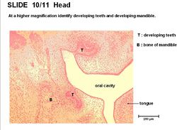
| − | [[Image:Tooth Development Histology.jpg|thumb|right|250px|Histología del Diente - Copyright RVC 2008]] | + | [[Image:Tooth Development Histology.jpg|thumb|right|250px|Tooth Histología del Diente - Copyright RVC 2008]] |
El desarrollo del diente se produce en las siguientes fases; | El desarrollo del diente se produce en las siguientes fases; | ||
| − | 1. Engrosamiento focal del epitelio oral en la cara medial del [[Gingiva#Surco Labiogingival|surco labiogingival]] forma la lámina dental. | + | 1. Focal thickening of oral epithelium on the medial aspect of the [[Gingiva#Surco Labiogingival|surco labiogingival]] forms the dental lamina. |
| − | + | Engrosamiento focal del epitelio oral en la cara medial del [[Gingiva#Surco Labiogingival|surco labiogingival]] forma la lámina dental. | |
2. El mesénquima se condensa en cada lámina. | 2. El mesénquima se condensa en cada lámina. | ||
3. La lámina dental se invagina para formar el brote dental. | 3. La lámina dental se invagina para formar el brote dental. | ||
| + | 4. El brote se expande y ramas dentales para convertirse en el órgano del esmalte. | ||
| + | 5. El órgano del esmalte rodea la células de la cresta neural derivada, papila dental. | ||
| + | 6. La combinación del órgano del esmalte y la papila dental constituye el diente temporal. | ||
| + | 7. pequeña masa de células de la yema de la lámina dental que forma el primordio del diente permanente que continúa el desarrollo. | ||
| + | 8. La capa celular interna del órgano del esmalte (del epitelio oral) se diferencia en ameloblastos . | ||
| + | 9. las células vecinas en las papilas dentales (a partir de células de la cresta neural) se diferencian en odontoblastos . | ||
| + | 10. La dentina rodea la pulpa para producir la raíz del diente. | ||
| + | 11. Las células epiteliales cerca de la forma del diente distal cementoblastos , secretoras de cemento alrededor de los dientes de raíz . | ||
| + | Hay una inducción interacción recíproca entre el epitelio oral y precursores mesénquima. El mesénquima forma del diente, que tiene propiedades diferenciativa lábil, pero estable propiedades morfogénica. formación de los dientes se inicia en la corona y progresa hacia la raíz . El diente no adquiere toda su longitud hasta que la corona se ha convertido. crecimiento de los dientes es aposición. | ||
| + | 3. The dental lamina invaginates to form the dental bud. | ||
| − | 4. | + | 4. The dental bud expands and branches to become the enamel organ. |
| − | 5. | + | 5. The enamel organ surrounds the neural crest cell derived, dental papilla. |
| − | 6. | + | 6. The combination of the enamel organ and dental papillae forms the deciduous tooth. |
| − | 7. | + | 7. Small mass of cells bud off the dental lamina forming the primordium of the permanent tooth which continues development. |
| − | 8. | + | 8. The inner cell layer of enamel organ (from oral epithelium) differentiates into [[Enamel Organ#Ameloblasts|ameloblasts]]. |
| − | 9. | + | 9. Neighbouring cells in the dental papillae (from neural crest cells) differentiate into [[Enamel Organ#Odontoblasts|odontoblasts]]. |
| − | 10. | + | 10. Dentine surrounds [[Enamel Organ#Pulp|pulp]] to produce the [[Enamel Organ#Root|root]] of the tooth. |
| − | 11. | + | 11. Epithelial cells near the distal tooth form [[Enamel Organ#Cementoblasts|cementoblasts]], secreting [[Enamel Organ#Cementum|cementum]] around the tooth [[Enamel Organ#Root|root]]. |
| − | + | There is a reciprocal inductive interaction between the oral epithelium and mesenchyme precursors. The mesenchyme forms the tooth, it has labile differentiative properties but stable morphogenic properties. Tooth formation starts at the [[Enamel Organ#Crown|crown]] and progresses towards the [[Enamel Organ#Root|root]]. The tooth does not acquire full length until the crown has emerged. Tooth growth is appositional. | |
| − | == | + | ==Eruption== |
| − | === | + | ===Deciduous Tooth=== |
| − | + | Eruption occurs after the [[Enamel Organ#Crown|crown]] has fully formed (prior to complete [[Enamel Organ#Root|root]] formation). It provides the space required for [[Enamel Organ#Root|root]] completion. The epithelial covering is continuous with gums after eruption. Erosion (wear) removes the epithelium. The 'toothless' gene stops eruption. | |
| − | |||
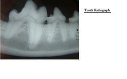
| − | === | + | [[Image:Tooth Radiograph.jpg|thumb|right|250px|Tooth Radiograph - Copyright Nottingham 2008]] |
| + | ===Permanent Tooth=== | ||
| − | + | The tooth migrates into the socket of the deciduous tooth on the lingual side. It increases the pressure on the deciduous tooth by increased growth. Resorption of the deciduous tooth root leads to its loosening. The deciduous tooth then sheds and the permanent tooth replaces it. Premature loss of the deciduous tooth leads to disorganised (non-occluding) permanent teeth. | |
==Enlaces== | ==Enlaces== | ||
| − | + | Click here for the [[Cavidad Oral - Anatomía & Fisiología - Flashcards#Teeth & Gingiva Flashcards|teeth & gingiva flashcards]]. | |
[[Categoría:Dientes - Anatomía & Fisiología]] | [[Categoría:Dientes - Anatomía & Fisiología]] | ||
Revisión del 21:34 9 may 2011
Introduction
Dientes se desarrollan de manera diferente en distintas regiones de la boca en la mayoría de las especies, un proceso llamado heterodonty. En algunos animales, los dientes se desarrollan de forma idéntica en las diferentes regiones de la boca, un proceso llamado homodonty. Las diferentes especies tienen diferentes números de dientes y de formas diferentes, dependiendo en gran parte de su dieta. No todas las especies poseen dientes y hay una gran variación en las fórmulas dentales entre las especies que tienen dientes. Los dientes se utilizan principalmente para la masticación - masticar y moler las partículas de alimentos, pero también se utilizan para agarrar la presa y para tirar. La superficie de oclusión se forma donde los dientes tocan. La superficie de contacto se forma donde los dientes adyacentes tocan.
Desarrollo del Diente
El desarrollo del diente se produce en las siguientes fases;
1. Focal thickening of oral epithelium on the medial aspect of the surco labiogingival forms the dental lamina. Engrosamiento focal del epitelio oral en la cara medial del surco labiogingival forma la lámina dental. 2. El mesénquima se condensa en cada lámina.
3. La lámina dental se invagina para formar el brote dental. 4. El brote se expande y ramas dentales para convertirse en el órgano del esmalte. 5. El órgano del esmalte rodea la células de la cresta neural derivada, papila dental. 6. La combinación del órgano del esmalte y la papila dental constituye el diente temporal. 7. pequeña masa de células de la yema de la lámina dental que forma el primordio del diente permanente que continúa el desarrollo. 8. La capa celular interna del órgano del esmalte (del epitelio oral) se diferencia en ameloblastos . 9. las células vecinas en las papilas dentales (a partir de células de la cresta neural) se diferencian en odontoblastos . 10. La dentina rodea la pulpa para producir la raíz del diente. 11. Las células epiteliales cerca de la forma del diente distal cementoblastos , secretoras de cemento alrededor de los dientes de raíz . Hay una inducción interacción recíproca entre el epitelio oral y precursores mesénquima. El mesénquima forma del diente, que tiene propiedades diferenciativa lábil, pero estable propiedades morfogénica. formación de los dientes se inicia en la corona y progresa hacia la raíz . El diente no adquiere toda su longitud hasta que la corona se ha convertido. crecimiento de los dientes es aposición. 3. The dental lamina invaginates to form the dental bud.
4. The dental bud expands and branches to become the enamel organ.
5. The enamel organ surrounds the neural crest cell derived, dental papilla.
6. The combination of the enamel organ and dental papillae forms the deciduous tooth.
7. Small mass of cells bud off the dental lamina forming the primordium of the permanent tooth which continues development.
8. The inner cell layer of enamel organ (from oral epithelium) differentiates into ameloblasts.
9. Neighbouring cells in the dental papillae (from neural crest cells) differentiate into odontoblasts.
10. Dentine surrounds pulp to produce the root of the tooth.
11. Epithelial cells near the distal tooth form cementoblasts, secreting cementum around the tooth root.
There is a reciprocal inductive interaction between the oral epithelium and mesenchyme precursors. The mesenchyme forms the tooth, it has labile differentiative properties but stable morphogenic properties. Tooth formation starts at the crown and progresses towards the root. The tooth does not acquire full length until the crown has emerged. Tooth growth is appositional.
Eruption
Deciduous Tooth
Eruption occurs after the crown has fully formed (prior to complete root formation). It provides the space required for root completion. The epithelial covering is continuous with gums after eruption. Erosion (wear) removes the epithelium. The 'toothless' gene stops eruption.
Permanent Tooth
The tooth migrates into the socket of the deciduous tooth on the lingual side. It increases the pressure on the deciduous tooth by increased growth. Resorption of the deciduous tooth root leads to its loosening. The deciduous tooth then sheds and the permanent tooth replaces it. Premature loss of the deciduous tooth leads to disorganised (non-occluding) permanent teeth.
Enlaces
Click here for the teeth & gingiva flashcards.


