Diferencia entre revisiones de «Organo de Esmalte»
(Página creada con '==Anatomy of the Enamel Organ== thumb|right|150px|Histology of Enamel Organ - Copyright RVC 2008 [[Image:Enamel Organ Layers.jpg|thu...') |
|||
| Línea 1: | Línea 1: | ||
| − | == | + | ==Anatomía del Organo de Esmalte== |
| − | [[Image:Soft Histology of Enamel Organ.jpg|thumb|right|150px| | + | [[Image:Soft Histology of Enamel Organ.jpg|thumb|right|150px|Histología del Organo de Esmalte - Copyright RVC 2008]] |
| − | [[Image:Enamel Organ Layers.jpg|thumb|right|150px| | + | [[Image:Enamel Organ Layers.jpg|thumb|right|150px|Capas del Organo de Esmalte - Copyright RVC 2008]] |
| − | [[Image:Thomes' Fibres Histology.jpg|thumb|right|150px|Thomes' | + | [[Image:Thomes' Fibres Histology.jpg|thumb|right|150px|Fibras Thomes' - Copywright RVC 2008]] |
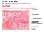
The main components which form the enamel organ are: | The main components which form the enamel organ are: | ||
*'''Outer epithelium''' | *'''Outer epithelium''' | ||
*'''Stellate reticulum'''- star shaped cells lying between the outer and inner epithelial layers. It has the appearance of connective tissue but is of epithelial derivation. | *'''Stellate reticulum'''- star shaped cells lying between the outer and inner epithelial layers. It has the appearance of connective tissue but is of epithelial derivation. | ||
| − | *'''Inner epithelium''' which becomes the enamel secreting [[ | + | *'''Inner epithelium''' which becomes the enamel secreting [[Organo de Esmalte#Ameloblastos|ameloblasto]] layer |
==Componentes== | ==Componentes== | ||
| Línea 15: | Línea 15: | ||
The crown of incisors have only one '''cusp'''. The crown of molars have up to 4 cusps for the grinding of food. | The crown of incisors have only one '''cusp'''. The crown of molars have up to 4 cusps for the grinding of food. | ||
| − | === | + | ===Raíz=== |
| − | Teeth may have one or more roots. The furcation angle is the point where roots diverge. The root ends in an apex which is where the nerves, blood vessels and lymphatics travel to the [[ | + | Teeth may have one or more roots. The furcation angle is the point where roots diverge. The root ends in an apex which is where the nerves, blood vessels and lymphatics travel to the [[Organo de Esmalte#Pulpa Dental|pulpa dental]]. '''Hypsodont''' teeth can have open roots (aradicular) e.g. in rabbits which have continued growth. Hypsodont teeth can have closed roots (radicular) e.g. horse where growth decreases with age. '''Brachydont''' teeth have no capacity for growth and so the roots are closed. |
'''Diferencias Entre las Especies''' | '''Diferencias Entre las Especies''' | ||
| − | The apex has a single foramen in dogs and cats. It remains open in herbivores. In the horse, the apex closes as the animal ages. Brachiocephalic dogs often have fused roots. Equine incisors have fused roots. In the horse's canines, the size of the root is much larger than the [[ | + | The apex has a single foramen in dogs and cats. It remains open in herbivores. In the horse, the apex closes as the animal ages. Brachiocephalic dogs often have fused roots. Equine incisors have fused roots. In the horse's canines, the size of the root is much larger than the [[Organo de Esmalte#Corona|corona]]. |
| − | ===Alveolar | + | ===Hueso Alveolar=== |
| − | The alveolar processes of the jaw consists of the '''alveolar | + | The alveolar processes of the jaw consists of the '''hueso alveolar''', '''trabecular bone''' and '''compact bone'''. |
| − | The densest bone called the '''cribiform plate''' lines the alveolus. This appears white on radiographs and is referred to as the '''[[ | + | The densest bone called the '''cribiform plate''' lines the alveolus. This appears white on radiographs and is referred to as the '''[[Organo de Esmalte#Lamina Dura|lamina dura]]'''. |
===Lamina Dura=== | ===Lamina Dura=== | ||
| − | The '''lamina dura''' lines the [[ | + | The '''lamina dura''' lines the [[Organo de Esmalte#Hueso Alveolar|hueso alveolar]]. If uninterrupted, it indicates good dental health. |
The '''lamina dura''' is seen as a white line radiographically. | The '''lamina dura''' is seen as a white line radiographically. | ||
| − | === | + | ===Esmalte=== |
| − | Enamel has an '''ectodermal''' origin. It is synthesised by [[ | + | Enamel has an '''ectodermal''' origin. It is synthesised by [[Organo de Esmalte#Ameloblastos|ameloblastos]]. It is very hard, densly calcified and '''acellular''', therefore cannot regenerate. |
| − | Complicated enamel folding occurs in teeth where the '''[[ | + | Complicated enamel folding occurs in teeth where the '''[[Organo de Esmalte#Corona|coronas]]''' are high. Enamel forming secretions pass through processes of apical cytoplasmic extension called '''Thomes' Fibres'''. |
| − | === | + | ===Dentina=== |
| − | '''Dentine''' is a calcified, collagen rich matrix. It is synthesised by '''[[ | + | '''Dentine''' is a calcified, collagen rich matrix. It is synthesised by '''[[Organo de Esmalte#Odontoblastos|odontoblastos]]'''. |
'''Secondary dentine''' is produced throughout life and increases with rate of repair. It is darker in colour than '''primary dentine'''. | '''Secondary dentine''' is produced throughout life and increases with rate of repair. It is darker in colour than '''primary dentine'''. | ||
| − | === | + | ===Cemento=== |
| − | '''Cementum''' is synthesised by '''[[ | + | '''Cementum''' is synthesised by '''[[Organo de Esmalte#Cementoblastos|cementoblastos]]'''. It is calcified tissue and lacks regular organisation. Collagen fibres extend from the cementum into the '''[[Organo de Esmalte#Ligamento Periodontal|ligamento periodontal]]''' to fasten the tooth in its socket. '''Cementum''' is relatively immune to pressure erosion, therefore the tooth can be be romedelled in its socket. |
| − | === | + | ===Pulpa Dental=== |
| − | Pulp fills the dental cavity. It is a delicate connective tissue bordering the [[ | + | Pulp fills the dental cavity. It is a delicate connective tissue bordering the [[Organo de Esmalte#Odontoblastos|odontoblasto]] layer. It is highly vascularised and contains a lymphatic plexus. |
Pulp allows pain sensation to thermal, mechanical and chemical stimulants. Most of the nervous supply is sensory, with some vasomotor input. | Pulp allows pain sensation to thermal, mechanical and chemical stimulants. Most of the nervous supply is sensory, with some vasomotor input. | ||
| − | ===Periodontal | + | ===Ligamento Periodontal=== |
| − | The collagen fibre bundles are called '''Sharpey's fibres'''. The fibres insert into the [[ | + | The collagen fibre bundles are called '''Sharpey's fibres'''. The fibres insert into the [[Organo de Esmalte#Hueso alveolar|hueso alveolar]] and [[Organo de Esmalte#Cemento|cemento]] of the tooth. |
There are 3 categories: gingival, trans-septal and alveolodental. There are evenly distributed blood vessels and nerve fibres transmitting thermal, pain and pressure sensation. Some species can also sense proprioception in the periodontal ligament. | There are 3 categories: gingival, trans-septal and alveolodental. There are evenly distributed blood vessels and nerve fibres transmitting thermal, pain and pressure sensation. Some species can also sense proprioception in the periodontal ligament. | ||
| Línea 57: | Línea 57: | ||
==Main Cells== | ==Main Cells== | ||
| − | [[Image:Ameloblast Histology.jpg|thumb|right|150px| | + | [[Image:Ameloblast Histology.jpg|thumb|right|150px|Histología de Ameloblasto - Copywright RVC 2008]] |
| − | === | + | ===Ameloblastos=== |
| − | '''Ameloblasts''' are cells in the enamel organ which forms the tooth. They secrete '''[[ | + | '''Ameloblasts''' are cells in the enamel organ which forms the tooth. They secrete '''[[Organo de Esmalte#Esmalte|esmalte]]'''. |
| − | Epithelial cells line the inner surface of the enamel organ. '''Ameloblasts''' are derived from epithelium and form a single layer of very long columnar cells that are hexagonal in cross section. They have elongated, basally sited nuclei. They synthesise '''[[ | + | Epithelial cells line the inner surface of the enamel organ. '''Ameloblasts''' are derived from epithelium and form a single layer of very long columnar cells that are hexagonal in cross section. They have elongated, basally sited nuclei. They synthesise '''[[Organo de Esmalte#Esmalte|esmalte]]''' which forms the '''[[Organo de Esmalte#Corona|corona]]''' of each tooth. They maintain connections with the newly synthesised '''[[Organo de Esmalte#Esmalte|esmalte]]''' through cellular projections called '''Thomes' fibres'''. |
| − | '''[[ | + | '''[[Organo de Esmalte#Esmalte|Esmalte]]''' is acellular so once the connection with the ameloblasts via the '''Thomes' fibres''' is lost (upon eruption), the [[Organo de Esmalte#Esmalte|esmalte]] matrix cannot be remodelled. |
| − | === | + | ===Odontoblastos=== |
| − | The '''odontoblasts''' are cells in the '''enamel organ''' which forms the tooth. They secrete '''[[ | + | The '''odontoblasts''' are cells in the '''enamel organ''' which forms the tooth. They secrete '''[[Organo de Esmalte#Dentina|dentina]]'''. |
| − | '''Odontoblasts''' are derived from mesenchyme and are composed of a single layer of elongated columnar cells. They are at the '''dental-pulp border'''. They secrete '''[[ | + | '''Odontoblasts''' are derived from mesenchyme and are composed of a single layer of elongated columnar cells. They are at the '''dental-pulp border'''. They secrete '''[[Organo de Esmalte#Dentina|dentina]]''' which is a mineralised matrix of collagen I, [[Organo de Esmalte#Dentina|dentina]] and proteins. |
| − | The first layer of [[ | + | The first layer of [[Organo de Esmalte#Dentina|dentina]] is formed on the enamel organ. As production increases, the odontoblasts are displaced from the [[Organo de Esmalte#Esmalte|esmalte]]. It is a major part of the tooth structure and is produced continually by the odontoblasts. The rate of [[Organo de Esmalte#Dentina|dentina]] synthesis is increased during repair as it is innervated (but still acellular). |
| − | === | + | ===Cementoblastos === |
| − | '''Cementoblasts''' are cells in the '''[[ | + | '''Cementoblasts''' are cells in the '''[[Organo de Esmalte#Esmalte|esmalte]]''' organ which forms the tooth. They secrete '''[[Organo de Esmalte#Cemento|cemento]]'''. |
| − | Epithelial cells are present near the distal end of the cup. They become follicle cells. '''Cementoblasts''' synthesise [[ | + | Epithelial cells are present near the distal end of the cup. They become follicle cells. '''Cementoblasts''' synthesise [[Organo de Esmalte#Cemento|cemento]] which mostly contains '''collagen I'''. |
| − | [[ | + | [[Organo de Esmalte#Cemento|Cemento]] surrounds the '''[[Organo de Esmalte#Dentina|dentina]]''' of the [[Organo de Esmalte#Raíz|raíz]]. [[Organo de Esmalte#Cemento|Cemento]] is acellular and not readily absorbed. |
| − | == | + | ==Comprobar tus Conocimientos == |
| − | [[Cavidad Oral - Anatomía & Fisiología_-_Flashcards# | + | [[Cavidad Oral - Anatomía & Fisiología_-_Flashcards#Dientes y Gingiva_Flashcards|Dientes y Gingiva - Flashcards]] |
[[Categoría:Dientes - Anatomía & Fisiología]] | [[Categoría:Dientes - Anatomía & Fisiología]] | ||
Revisión del 10:35 10 may 2011
Anatomía del Organo de Esmalte
The main components which form the enamel organ are:
- Outer epithelium
- Stellate reticulum- star shaped cells lying between the outer and inner epithelial layers. It has the appearance of connective tissue but is of epithelial derivation.
- Inner epithelium which becomes the enamel secreting ameloblasto layer
Componentes
The enamel organ has many different components. These consist of:
Corona
The crown is covered by enamel. It meets the root at the cemento-enamel junction (CEJ).
The crown of incisors have only one cusp. The crown of molars have up to 4 cusps for the grinding of food.
Raíz
Teeth may have one or more roots. The furcation angle is the point where roots diverge. The root ends in an apex which is where the nerves, blood vessels and lymphatics travel to the pulpa dental. Hypsodont teeth can have open roots (aradicular) e.g. in rabbits which have continued growth. Hypsodont teeth can have closed roots (radicular) e.g. horse where growth decreases with age. Brachydont teeth have no capacity for growth and so the roots are closed.
Diferencias Entre las Especies
The apex has a single foramen in dogs and cats. It remains open in herbivores. In the horse, the apex closes as the animal ages. Brachiocephalic dogs often have fused roots. Equine incisors have fused roots. In the horse's canines, the size of the root is much larger than the corona.
Hueso Alveolar
The alveolar processes of the jaw consists of the hueso alveolar, trabecular bone and compact bone.
The densest bone called the cribiform plate lines the alveolus. This appears white on radiographs and is referred to as the lamina dura.
Lamina Dura
The lamina dura lines the hueso alveolar. If uninterrupted, it indicates good dental health.
The lamina dura is seen as a white line radiographically.
Esmalte
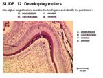
Enamel has an ectodermal origin. It is synthesised by ameloblastos. It is very hard, densly calcified and acellular, therefore cannot regenerate.
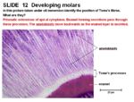
Complicated enamel folding occurs in teeth where the coronas are high. Enamel forming secretions pass through processes of apical cytoplasmic extension called Thomes' Fibres.
Dentina
Dentine is a calcified, collagen rich matrix. It is synthesised by odontoblastos.
Secondary dentine is produced throughout life and increases with rate of repair. It is darker in colour than primary dentine.
Cemento
Cementum is synthesised by cementoblastos. It is calcified tissue and lacks regular organisation. Collagen fibres extend from the cementum into the ligamento periodontal to fasten the tooth in its socket. Cementum is relatively immune to pressure erosion, therefore the tooth can be be romedelled in its socket.
Pulpa Dental
Pulp fills the dental cavity. It is a delicate connective tissue bordering the odontoblasto layer. It is highly vascularised and contains a lymphatic plexus.
Pulp allows pain sensation to thermal, mechanical and chemical stimulants. Most of the nervous supply is sensory, with some vasomotor input.
Ligamento Periodontal
The collagen fibre bundles are called Sharpey's fibres. The fibres insert into the hueso alveolar and cemento of the tooth.
There are 3 categories: gingival, trans-septal and alveolodental. There are evenly distributed blood vessels and nerve fibres transmitting thermal, pain and pressure sensation. Some species can also sense proprioception in the periodontal ligament.
Main Cells
Ameloblastos
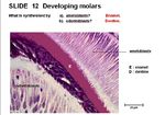
Ameloblasts are cells in the enamel organ which forms the tooth. They secrete esmalte.
Epithelial cells line the inner surface of the enamel organ. Ameloblasts are derived from epithelium and form a single layer of very long columnar cells that are hexagonal in cross section. They have elongated, basally sited nuclei. They synthesise esmalte which forms the corona of each tooth. They maintain connections with the newly synthesised esmalte through cellular projections called Thomes' fibres.
Esmalte is acellular so once the connection with the ameloblasts via the Thomes' fibres is lost (upon eruption), the esmalte matrix cannot be remodelled.
Odontoblastos
The odontoblasts are cells in the enamel organ which forms the tooth. They secrete dentina.
Odontoblasts are derived from mesenchyme and are composed of a single layer of elongated columnar cells. They are at the dental-pulp border. They secrete dentina which is a mineralised matrix of collagen I, dentina and proteins.
The first layer of dentina is formed on the enamel organ. As production increases, the odontoblasts are displaced from the esmalte. It is a major part of the tooth structure and is produced continually by the odontoblasts. The rate of dentina synthesis is increased during repair as it is innervated (but still acellular).
Cementoblastos
Cementoblasts are cells in the esmalte organ which forms the tooth. They secrete cemento.
Epithelial cells are present near the distal end of the cup. They become follicle cells. Cementoblasts synthesise cemento which mostly contains collagen I.
Cemento surrounds the dentina of the raíz. Cemento is acellular and not readily absorbed.


