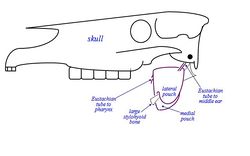
Bolsas Guturales - Anatomía & Fisiología
Also known as: Auditory Tube Diverticulum
Introducción
The Guttural Pouch is present only in members of the order Perissodactyla (nonruminant ungulates: horses, tapirs, rhinoceros) and another small band of small mammals including Hyraxes, certain bats and a South American mouse.
The guttural pouches are paired ventral diverticulae of the eustachian (auditory) tubes, formed by escape of mucosal lining of the tube through a relatively long ventral slit in the supporting cartilages. The auditory tube connect the nasal cavity and middle ear and the diverticulum dilates to form pouches which can have a capacity of 300-500ml in the domestic horse. The pouches are normally aair filled.
La bolsa gutural está presente sólo en los miembros de la orden Perissodactyla (ungulados no rumiantes: caballos, tapires, rinocerontes) y otro pequeño grupo de pequeños mamíferos como damanes, algunos murciélagos y un ratón de América del Sur. Las bolsas guturales se aparean divertículos ventral del Eustaquio (auditiva), tubos, formado por el escape de la mucosa del tubo a través de una hendidura ventral relativamente largo en los cartílagos de soporte. La trompa de Eustaquio conecta la cavidad nasal y del oído medio y el divertículo se dilata para formar bolsas que pueden tener una capacidad de 300-500ml en el caballo doméstico. Las bolsas son normalmente AAIR llena.
Estructura
The Guttural Pouch is located below the cranial cavity, towards the caudal end of the skull/wing of atlas. It is covered laterally by the Pterygoid muscles, parotid and mandibular glands. The floor lies mainly on the pharynx and beginning of the Oesophagus. The medial retropharyngeal lymph node lies between the pharynx and ventral wall of the pouches.
Right and left pouches are separated dorsomedially by rectus capitis ventralis and longus capitis muscles. Below this, by fused walls of the two pouches, the median septum is formed.
Each pouch is moulded to the stylohyoid muscle which divides the medial and lateral compartments, the medial compartment being approximately double the size of the lateral one and extends further caudally and ventrally.
The guttural pouch has close association with many major structures including several cranial nerves (glossopharyngeal, vagus, accessory, hypoglossal), the sympathetic trunk and the external and internal carotid arteries. The pouch directly covers the temporohyoid joint. The pouch has an extremely thin wall which is lined by respiratory epithelium which secretes mucus. This normally drains into the pharynx when the horse is grazing.
Several cranial nerves and arteries lie directly against the pouch as they pass to and from foramina in the caudal part of the skull (vessels within mucosal folds that indent the pouches):
Medial Compartment:
- Cranial nerves IX, X, XI, XII.
- Continuation of the sympathetic trunk beyond the cranial cervical ganglion.
- Internal carotid artery.
Lateral Compartment:
- Cranial nerve VII - limited contact with the dorsal part of the compartment.
- External carotid artery crosses the lateral wall of the lateral compartment in its approach (as maxillary artery) to the atlas canal. The external maxillary vein is also visible.
Drainage:
Natural drainage of the pouch is throught the slit-like (pharyngeal) openings of the eustachian tube in the lateral wall of the nasopharynx. The connection opens when the horse swallows and grazing normally provides drainage. However, most of the pouch is ventral to his slit, and therefore drainage may be rather ineffective. If blocked, secretions accumulate and the pouch distends producing a palpable swelling.
La bolsa gutural se encuentra por debajo de la cavidad craneal, hacia el extremo caudal del cráneo / ala del atlas. Está cubierta lateralmente por los músculos pterigoideo, parótida y mandibular glándulas. La planta se encuentra principalmente en la faringe y principios del esófago . Los ganglios linfáticos retrofaríngeos medial se encuentra entre la faringe y la pared ventral de las bolsas. Derecha y la izquierda bolsas se separan dorsomedialmente por capitis ventralis recto y los músculos del longus capitis. Por debajo de este, por las paredes fundido de los dos sobres, el septo medio se forma. Cada bolsa se moldea al músculo estilohioideo que divide los compartimentos medial y lateral, el compartimento medial que es aproximadamente el doble del tamaño de la lateral y se extiende más caudal y ventral. La bolsa gutural tiene estrecha relación con muchas de las estructuras más importantes, incluyendo varios nervios craneales (glosofaríngeo, vago, accesorio, hipogloso), el tronco simpático y la arteria carótida interna y externa. La bolsa cubre directamente la articulación temporohyoid. La bolsa tiene una pared delgada que es extremadamente revestidos por epitelio respiratorio que segrega moco. Esto normalmente desemboca en la faringe cuando el caballo es el pastoreo. Varios pares craneales y las arterias se encuentran directamente en contra de la bolsa a medida que pasan hacia y desde agujeros en la parte caudal del cráneo (los buques dentro de pliegues de la mucosa que las bolsas guión): Compartimento medial : Los nervios craneales IX, X, XI, XII. Continuación del tronco simpático más allá del ganglio cervical craneal. Arteria carótida interna. Compartimiento lateral : Los nervios craneales VII - un contacto limitado con la parte dorsal del compartimiento. la arteria carótida externa atraviesa la pared lateral del compartimento lateral en su enfoque (como la arteria maxilar) al canal atlas. La vena maxilar externa también es visible.
Drenaje: drenaje natural de la bolsa se cree la raja-como (la faringe) las aberturas de la trompa de Eustaquio en la pared lateral de la nasofaringe. La conexión se abre cuando las golondrinas y el pastoreo de caballos normalmente proporciona drenaje. Sin embargo, la mayor parte de la bolsa ventral es a su raja, y por lo tanto el drenaje puede ser bastante ineficaz. Si esto ocurriera, las secreciones se acumulan y la bolsa se distiende produciendo una hinchazón palpable.
Función
The function of guttural pouches is largely unknown, however hypotheses have been put forward:
- It may influence internal carotid artery blood pressure. Air pressure varies with phase and forcefulness of respiration and the artery in the mucosal fold is exposed sufficiently to be affected.
- Cerebral blood cooling mechanism. Operates at times of physical stress/exercise, when core body temperature is raised. Vigorous respiration, cool air in guttural pouches, and the exposed artery all lead to cooling of the blood.
Función
La función de las bolsas guturales es en gran parte desconocida, sin embargo, las hipótesis se han propuesto: Se puede influir en la presión interna sanguíneo de la arteria carótida. La presión del aire varía con la fase y la contundencia de la respiración y la arteria en la mucosa veces está expuesto lo suficiente como para ser afectado. sanguíneo cerebral mecanismo de enfriamiento. Funciona en momentos de estrés físico o ejercicio, cuando la temperatura central del cuerpo se eleva. respiración vigorosa, de aire fresco en las bolsas guturales, y la arteria expuestos todos conducen a un enfriamiento de la sangre.
Enlaces
Referencias
Dyce, K.M., Sack, W.O. and Wensing, C.J.G. (2002) Textbook of Veterinary Anatomy. 3rd ed. Philadelphia: Saunders.
