Retículo - Anatomía & Fisiología
Introducción
The reticulum is the second chamber of the ruminant stomach. It has regular contractions which precede the biphasic ruminal contraction for digestion of food particles. Mechanical digestion and microbial fermentation occur to breakdown food particles for absorption. Volatile fatty acids are the major product of ruminant digestion.
Estructura
The reticulum is covered by greater omentum. The rumino-reticular fold often gets objects lodged. When the rumen contracts, the object can be pushed through the reticulum wall into the pericardium and heart.
Opening at the cardia into both the reticulum and the rumen is called the reticular groove (see oseophageal groove). The reticular groove also opens into the omaso.
The reticulum is cranial to the rumen at ribs 6-8. It is located from cardia to the diaphragm. It lies above the xiphoid process of the sternum. Serosa covers the surface.
Función
The functions of the reticulum include waste removal and movement. Simpler products of digestion are assimilated directly, others continue down the digestive tract for further digestion.
La Contracción Ruminoreticular
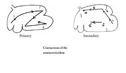
The contractions have to main functions:
- Primary contraction mixes food by a ruminoreticuluar mixing cycle. There are 2 contractions of the reticulum (2nd most powerful) which continue over the rumen. Ingesta flows from the reticulum to cranial rumenal sac and then to reticulum (or ventral sac). It occurs every 60 seconds.
- The secondary contraction lets gas out (see eructo). Ingesta flows from the ventral blind sac to the dorsal blind sac then to dorsal sac (eructation) and to the ventral sac.
Vascularización
The reticulum receives blood supply from the cranial mesenteric artery, celiac artery and right and left ruminal arteries.
Inervación
The reticulum is innervated by the dorsal vagus (CN X) (most important) and the ventral vagus nerve (CN X).
Linfáticos
Numerous small lymph nodes are scattered in the grooves. The lymph drains to larger atrial nodes between the cardia and omaso, then to the cistera chyli.
Histología
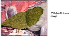
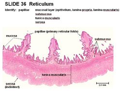
The reticulum is lined by a keratinised stratified squamous epithelium and there are no glands present. The characteristic honeycomb appearance is formed by the mucosal layer trown into short and tall folds. The folds gradually merge into papillae, where the reticulum meets the rumen. Conical papillae, also called secondary papillae, are present on folds, providing a rough surface to increase food breakdown.
The lamina muscularis is present as discrete bands of smooth muscle (not continuous). There are two thick layers of tunica muscularis, the inner circular and the outer longitudinal. The upper keratinised layer of the reticulum protects against abrasion and the deeper layers metabolise volatile fatty acids.
Diferencias Entre las Especies
Rumiantes Pequeños
Small ruminants have a larger reticulum compared to cattle. In sheep and goats, the ridges of the reticular cells are lower and have more prominent serrated edges than in cattle. The papillated ruminal mucosa expands over a greater proportion of the reticulum.
Enlaces
Haz "clic" aquí para información sobre el rumen
Haz "clic" aquí para información sobre el omaso
Haz "clic" aquí para información sobre el abomaso
| Retículo - Anatomía & Fisiología Entorno de Enseñanza Virtual | |
|---|---|
 Selección de videos relevantes |
Lateral view of the Abdomen of a young Ruminant Sections of the Ruminant Stomach Left sided topography of the Ovine Abdomen and Thorax Right sided topography of the Ovine Abdomen Structure of the ruminant forestomachs |


