Linfoma
También conocido como: Linfosarcoma — Malignant Lymphoma
Introducción
Lymphoma is caused by malignant clonal expansion of lymphoid cells and most commonly arises from lymphoid tissues including the bone marrow, thymus, lymph nodes and spleen. Lymphoma is documented to be the most common haematopoietic neoplasm in dogs.
Classification
- Cytological classification
- Well differentiaed (lymphocytic) - The malignant cells represent normal lymphocytes, although in excessive numbers.
- Poorly differentiated (lymphoblastic) - The malignant cells represent atypical lymphocytic cells with lymphoblastic characteristics.
- Tumour distribution
- Nodular/ follicular - A well organised pattern of slow growth, no metastasis, they are of the B-lymphocyte type
- Diffuse - Result in effacement of normal lymphoid architecture by a very homogeneous population of lymphoid cells.
- Anatomical classification
- Thymic - Only the timo is affected.
- Alimentary - Gut and associated lymphoid tissue affected.
- Multicentric - Widespread involvement of lymph nodes.
- Cutaneous lymphoma - Usually presents as generalised skin disease, but is a malignant transformation of T cells with a propensity for pithelial sites.
- Type of lymphocyte - T-cell, B-cel or NK-cell
- Time scale - Acute or Chronic
Order of prevalence in UK is cats, dogs, cattle, pigs and sheep. In the cat and ox, viral agents have been identified as the causal agents.
Perro
Lymphoma is one of the prevalent neoplasms in the dog. The incidence is about 28 per 100,000 dogs. Blood of affected dogs shows neither a relative nor absolute increase in the number of lymphocytes until the late stages of the disease. When this stage is reached, poorly differentiated cells may appear in the blood.
In the dog, multicentric lymphoma is most common representing 80% of cases. Alimentary, cutaneous, mediastinal and extranodal sites are less common. Additionally the majority of lymphoma cases in dogs are of the B-cell immunophenotype.
Gato
FeLV is an important cause of lymphoma in the cat. Following the introduction of widespread FeLV testing and vaccination the most common type of lymphoma affecting cats is alimentary when previously it had been mediastinal and multicentric forms. Only 10% of lymphoma cases in cats are now associated with FeLV, whereas it used to be 70%.
The alimentary form affects the mesenteric lymph nodes, intestine, hígado, spleen , and potentially the kidney. The thymic form presents as a thymic mass, also in the mediastinal lymph nodes. The pleural lymph nodes and the liver may potentially be affected. Multicentric form is found in the peripheral and deep lymph nodes, liver and spleen. The kidney may sometimes be affected. There is also a renal form affecting kidneys and other abdominal organs and leukaemic form affecting the bone marrow alone, this form is rare.
Horse
In horses, lymphoma is the most common haemopoietic neoplasm. It has been characterised into four main forms: alimentary, cutaneous, mediastinal and multicentric, however, it takes mainly the alimentary form.
Cattle
Cattle suffer both lymphosarcoma and leukosis in a variety of cytological forms. Bovine lymphoma is caused by Bovine Leukaemia Virus (BLV). There is a juvenile form of bovine lymphoma seen in young cattle which is not associated with BLV.
Cerdo
Porcine disease is mainly multicentric affecting lymph nodes, liver and spleen.
Oveja
Ovine lymphoma is uncommon. It may be multicentric or thymic.
Razas Más Afectadas
Perro
Affected dogs have a wide age range, most are middle-aged however young animals can be affected, 80% of cases affect the 5 to 11 year old age group. There may also be a male predilection.
Gato
The median age of affected cats is 9-10 years and oriental cat breeds may be predisposed.
Caballo
There are no sex, age or breed predilections.
Signos Clínicos
Perros
Multicentric Lymphoma
- The most common presenting sign in dogs is a lymphadenopathy, with only 10-20% of dogs presenting clinically unwell. Dogs that do present with clinical signs may be anorexic, lethargic and have lost weight.
For other types of lymphoma affecting dogs the clinical signs will demonstrate the anatomical site affected.
Mediastinal forms will present with dyspnoea due to compression of the trachea and upper respiratory tract. Dysphagia may also be present due to compression of the oespohagus. Dogs with mediastinal lymphoma can also have pitting oedema of the head and neck due to compression of the cranial vena cava. On ausculatation there is often an absence of lung sounds cranially and caudal displacement of the normal cardiac sounds, and dullness on percussion of the cranial thorax. Polyuria and polydypsia may be present due to paraneoplastic hyperlcalcaemia. Differential diagnoses for a cranial mediastinal mass are: thymoma, thyroid adenocarcinoma, a mediastinal abscess, or a branchial cyst.
Alimentary forms will present with signs of obstruction such as vomiting, diarrhoea, anorexia and thickened loops of intestine on abdominal palpation.
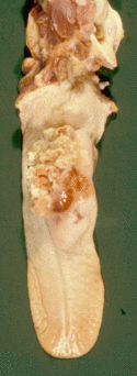
Cutaneous lymphoma can also occur with a varied presentation but often present as cutaneous nodules.
Gatos
In contrast to dogs, cats are more likely to present unwell. Again the clinical signs will depend on the anatomical location affected.
Alimentary cats will present with vomiting, diarrhoea, weight loss and anorexia.
Mediastinal cats will present with signs of compression of structures in the cranial thorax. These include dyspnoea, coughing and tachypnoea due to compression of the trachea. Weight loss and regurgitation may also occur secondary to compression of the oesophagus. On auscultation lung sounds are displaced caudally and lung sounds are decreased ventrally. There may be a loss of compressibility ('rib spring') over the cranial thorax. There may be pleural effusion present. Differential diagnoses for a cranial mediastinal mass are: thymoma, thyroid adenocarcinoma, a mediastinal abscess, or a branchial cyst.
Renal lymphoma also occurs in cats and affected animals will present with signs similar to renal failure.
Nasal lymphoma cases will present with dyspnoea, nasal discharge, epistaxis, facial pain or distortion and loss of airflow.
Caballo
A thoracic effusion may occur in the alimentary and multicentric forms of the disease, which usually has the characteristics of a modified transudate.
Mediastinal lymphoma also produces clinical signs such as pointing of the forelimb, tachycardia, distension of the jugular vein and caudal displacement of the heart - it may be confused with colic. It should be differentiated from mediastinal abscessation by ultrasound of the mass and cytology of pleural fluid.
Intra-abdominal neoplasia (which can be multicentric or alimentary) may presents with a history of chronic weight loss and inappetance, recurring colic and intermittant pyrexia.
Physical Examination
Gato y Perro
An abdominal mass may be palpable and bowel loops may feel thickened in alimentary lymphoma. Additionally enlarged mesenteric lymph nodes and enlarged abdominal organs may be palpable. Muffled heart sounds and a non-compressible thoracic region may be found in mediastinal lymphoma. Petechiae, anaemia and icterus may also be present in any form of lymphoma.
Caballo
Mediastinal masses can sometimes be palpable externally at the base of the jugular groove, due to the mass extending through the thoracic inlet.
Diagnosis
Laboratory Tests
Haematological analysis should always be performed with suspected lymphoma for staging purposes and for the recording of base-line parameters prior to the initiation of any treatment to assess the severity of any future myelosuppression. Potential abnormalities for those patients with bone marrow involvement may include lymphocytosis, thrombocytopenia, neutropenia and the presence of immature lymphoid precursors.
Affected cats are not usually leukemic.
On biochemistry abnormalities may include hypoproteinaemia, elevated hepatic enzymes and elevated Blood Urea Nitrogen /creatinine.
All cats with suspected lymphoma should be tested for FeLV and FIV, usually performed via enzyme-linked immunosorbent assay (ELISA) available in general practice in kit form (CITE test). Virus isolation would be required for a definitive result, however this is not only more time consuming but is more expensive. An ELISA is also frequently used for the diagnosis of FIV.
Paraneoplastic Syndrome Dogs may present with hypercalcaemia, this is due to the release of parathyroid hormone - related protein (PTHrp) released by the tumor, which produces these effects by acting like parathyroid hormone. Affected cats are not usually hypercalcaemic.
Radiography
A mass may be visible via plain or contrast abdominal radiography. Both abdominal and thoracic imaging is required in assessing the surrounding structures.
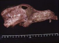
For nasal lymphoma, radiography of the head may reveal: increased soft tissue densities in the nasal cavities and possibly loss of turbinate structure.
Ultrasonography
Superior to radiography in assessing infiltration or abnormalities of tissue architecture and assessing the surrounding structures for metastasis. Guided aspirates or biopsies may also be taken at this time, including lymph node sampling, to evaluate degree of systemic involvement.
Cytology
Cytology is a necessary tool in the work-up of a lymphoma case. It provides both a diagnosis and a prognosis when combined with the entire clinical picture. Lymphoma produces a cell population which is both distinct and recognisable, allowing identification and classification of the type of lymphoma by cytology. Fine needle aspiration is a quick, cheap, non-invasive and effective method for collecting cells for cytology, and should always be considered a first-line test. Ideally cytology should always be supported by histology.
Cytology can also be used to examine pleural fluid samples if there is a suspicion of neoplasia.
Smears should be stained and examined microscopically.
Cytological criteria for lymphoma:
- Large amounts of lymphoblasts
- Large nuclei and prominent nucleoli
- High mitotic rate - bizarre mitotic figures may be present
- Small volume of basophilic cytoplasm
- Coarse chromatin
These features can be assessed to determine the grade of tumour and therefore the likely treatment response and progression of disease. Small well-differentiated lymphocytes normally suggest a low-grade lymphoma, and large, poorly differentiated lymphoid cells suggest a higher grade of lymphoma.
Perros
- Canine lymphoma is normally multicentric, therefore the ideal method for collecting a sample for cytological examination is fine needle aspiration of the lymph nodes. Ideally samples should come from multiple nodes to give a representative sample. Popliteal and prescapular lymph nodes are easily accessible and therefore ideal for sampling. Submandibular lymph nodes should be avoided where possible as they are commonly enlarged and reactive as a result of dental disease. It should be noted that canine lymphoma can occur in any organ containing lymphoid tissue.
Gatos
- Feline lymphoma is more variable in its presentation, with the three types (mediastinal, alimentary and multicentric) common in general practice. The sample taken for cytological examination should be appropriate for the type of lymphoma:
- Ultrasound guided aspirates, partial thickness endoscopic grab biopsies or full thickness biopsies via exploratory laparotomy for intenstinal lymphoma
- Pleural fluid aspirate with or without supporting ultrasounded-guided aspirate or core biopsy of the mass (which will differentiate it from thymoma) with mediastinal lymphoma
- Peripheral lymph node aspirates for multicentric lymphoma
- Lymphoma can occur in any tissue containing lymphoid tissue, for example the eye, kidney, CNS, liver, upper respiratory tract, lungs and skin. Cytology is an essential tool for diagnosis in these cases, as the lymphoma can present with variable clinical signs and diagnosis can only be confirmed using cytology. As mentioned above, the cytological diagnosis should be supported by histopathology if possible, particularly if the cytological sample is equivocal.
- NB. Lymphoma should not be confused with reactive lymphoid hyperplasia in the healthy cat. Generalised lymphadenopathy may present like multicentric lymphoma but is infact a natural immune response in the healthy cat. The same should be considered in other types of lymphoma, for example hepatic lymphoma looks cytologically identical to lymphocytic periportal hepatitis, and it is necessary to incorporate the entire clinical picture when making a diagnosis. Histopathological sampling is ideal for confirming the diagnosis.
Caballo
- In equine lymphoma, neoplastic cells are not always present, but when they are this may allow diagnosis.
- A sample of pleural or peritoneal fluid may be taken and examined cytologically if it is present. Otherwise a direct fine needle aspirate of the mass of lymph nodes may be performed. The fluid should be a modified transudate and contain a mixed cell population. Neoplastic lymphocytes are pleomorphic round cells that demonstrate anisocytosis and anisokaryosis and have very basophilic cytoplasm. If these cells are present then the diagnosis of lymphoma can be confirmed, otherwise surgical biopsy may be necessary.
Biopsy
A biopsy may be required if diagnosis cannot be made from FNA's. This may occur if; the aspirate provided a low number of cells; the cells were badly preserved; the disease is in its early stages or the neoplastic cells are small. If the lymph node is biopsied, it is best to remove the entire node in an excisional biopsy so the tissue architecture remains intact.
Biopsy may also be indicated it the neoplasia is localised to a specific organ which is not amenable to ultrasound guided FNA, for example the gastrointestinal tract.
Nasal lymphoma can be diagnosed by rhinoscopic or blind biopsy using a suction-catheter or grab-forceps technique.
Bone marrow aspiration or biopsy is needed to stage the disease.
Patología
Secondary liver tumours are the most common secondary malignancy. They can be present as nodules or as diffuse infiltration along the portal tracts. Grossly, the liver is enlarged, turgid and friable with many minute pale foci. The whole organ is diffusely pale. Microscopically, tumour cells are seen to spread diffusely through the sinusoids.
Splenomegaly occurs in multicentric lymphosarcoma. Splenic enlargement may be marked if any form of lymphosarcoma is in leukaemic phase.
Staging
A staging system is used for lymphoma (Owen, 1980):
- Stage I - Involvement limited to a single node or lymphoid tissue in a single organ (excluding bone marrow)
- Stage II - Involvement of many lymph nodes in a regional area (+/- tonsils)
- Stage III - Generalised lymph node involvement
- Stage IV - Liver and/or spleen involvement (+ stage III)
- Stage V - Manifestations in the blood and involvement of bone marrow and/or other organ systems (+/-stages I-IV)
Each stage is then subclassifed as a) without systemic signs or b) with systemic signs.
Tratmiento
Gatos y Perros
Surgery
- Firstly, a laparotomy is required for many cases of alimentary lymphoma to obtain biopsy material. For solitary masses without systemic disease resection and anastomosis of the intestine is advised (single modality treatment). Local resection in cats has occasionally been curative. Other focal lyphoma may also be resected, however surgery alone may be insufficient for long-term control of the disease and if not all the tumour is able to be resected. Should relapse occur, or if there is systemic progression, chemotherapy will be required (multimodal treatment).
Radiotherapy
- Lymphoma is highly radiosensitive and in theory radiotherapy should be efficient in treating all forms of lymphoma, however, surrounding tissues often have a low tolerance.
Chemotherapy
- Combination chemotherapy is the most frequent method of treatment and the most commonly used protocols include:
- COP which consists of Cyclophosphamide, Vincristine and Prednisolone. It is frequently used in cats and can be used for induction therapy (8 weeks) as well as a long term maintenance protocol.
- COAP consists of Cyclophosphamide, Vincristine, Prednisolone and Cytosine arabinoside
- CHOP consists of Cyclophosphamide, Vincristine, Prednisolone and Doxorubicin.
- Corticosteroids must not be administered prior to initiation of chemotherapy as they can cause resistance to cytotoxics and hence reduce the rate of response and the survival time. The aim is to induce remission and then continue with a maintenance regime, adjusting the dose as required with rescue therapy should relapse occur.
- Response to treatment can be monitored via reduction in tumour mass and size of lymph nodes. Haematological values should be frequently monitored to assess the effects of the drugs. In particular, animals should be monitored for the presence of azotaemia, neutropenia/sepsis, hypercalcaemia and pyrexia.
Supportive Therapy Whilst receiving chemotherapy. patients should receive a high quality, palatable diet to maintain calorific intake. If animals become anorexic they should receive appetite stimulation in cats e.g Cyproheptadine (Periactin) or antiemetics if vomiting occurs. Additionally, fluid therapy, laxatives and analgesia may be required.
Caballos
Treatment is symptomatic and euthanasia may be required with the progression of clinical signs.
Prognosis
Gatos y Perros
The mean survival times for dogs and cats without therapy is 6-8 weeks. For those receiving corticosteroids alone is 3 months.
If chemotherapy is administered then the mean survival time increases to 6-9 months. Local canine lymphoma responds better to chemotherapy than the diffuse form of disease. Immunophenotype (T cell versus B cell lymphoma) does not appear to be associated with prognosis in cats as it can be in dogs. Factors indicating a better prognosis (overall survival) in cats include: an early presentation, a complete initial response to treatment and a clinically well patient (‘substage a’ disease).
In cats, response rate to induction chemotherapy is 26-79% and there is an apparently a poorer response rate in cats compared with dogs, however, 30-40% of cats that do have complete remission and will maintain complete remission for two years or more and long-term maintenance chemotherapy can frequently be stopped and many will then live free of disease. Hence, dogs may have higher remission rates but are less likely than cats to be able to maintain remission without chemotherapy.
Caballo
The prognosis is poor and definitive diagnosis is usually achieved on post-mortem examination.
Referencias
Copas, V (2011) Diagnosis and treatment of equine pleuropneumonia In Practice 2011 33: 155-16
Cowell, R. (2002) Diagnostic cytology and haematology of the horse Elsevier Health Sciences
Freeman, KP (2007) Self-Assessment Colour Review of Veterinary Cytology - Dog, Cat, Horse and Cow Manson
Gear, R (2009) Practical update on canine lymphoma : 1. Classification and Diagnosis In Practice 2009 31: 380-384
Hayes A. (2006) Feline lymphoma 1. Principles of diagnosis and management, In Practice, 28, pp 516-524
Hayes, A (2006) Feline lymphoma 2. Specific Disease Presentations In Practice 2006 28, pp 578-585
Head K. W, Else R. W, Dubielzig R.R, (2002) Tumours of the Alimentary Tract, in Tumours in Domestic Animals, 4th edition, Ed Menten D. J, Iowa State Press, Blackwell Publishing, Iowa, pp 471-472
Hewetson, M (2006) Investigation of false colic in the horse In Practice 2006 28: 326-33
Milne, E (2004) Peritoneal fluid analysis for the differentiation of medical and surgical colic in horses In Practice 2004 26: 444-44
Morris J, Dobson J (2001) Gastrointestinal Tract, in Small Animal Oncology, Blackwell Science, pp 228-239
Selting K. A, (2007), Intestinal Tumours, Cancer of the Gastrointestinal Tract, in Withrow and MacEwen's Small Animal Clinical Oncology, fourth edition, Eds Withrow S.J, Vail D.M, Missouri, Saunders Elsevier, pp 491-501
Sparks, AH & Caney, SMA (2005) Self-Assessment Colour Review Feline Medicine Manson
Stell, A (2009) Haemopoetic Neoplasia - Lymphoreticular and Haemopoetic System RVC Intergrated BVetMed Course, Royal Veterinary College
White, R. A. S, (2003), Tumours of the intestines, in BSAVA Manual of Canine and Feline Oncology, second edition, Eds Dobson J. M, Lascelles B. D. X, Gloucester, British Small Animal Veterinary Association, pp 229-233
| Este artículo ha sido revisado por pares, pero aún no ha sido evaluado por un experto. |


