Diferencia entre revisiones de «Páncreas - Anatomía & Fisiología»
| (No se muestran 2 ediciones intermedias del mismo usuario) | |||
| Línea 15: | Línea 15: | ||
'''La Secreción Alcalina''' | '''La Secreción Alcalina''' | ||
| − | El jugo pancreático vierte en el duodeno a través de conductos. Es alcalina, ya que contiene '''bicarbonato''' y los '''iones de cloruro'''. Los iones de bicarbonato son transportados activamente al lumen del conducto. El agua sigue pasivamente por ósmosis. La osmolaridad del jugo pancreático es equivalente a la osmolaridad de la sangre. El jugo pancreático es alcalino para neutralizar el jugo gástrico ácido. Esto es ventajoso porque: proporciona un pH óptimo de las enzimas del páncreas y evita daños a la mucosa delgada de absorción del duodeno. Su alcalinidad también ayuda a amortiguar el [[Intestino Grueso - Anatomía & Fisiología|intestino grueso]] que es importante en [[Fermentadores del Intestino | + | El jugo pancreático vierte en el duodeno a través de conductos. Es alcalina, ya que contiene '''bicarbonato''' y los '''iones de cloruro'''. Los iones de bicarbonato son transportados activamente al lumen del conducto. El agua sigue pasivamente por ósmosis. La osmolaridad del jugo pancreático es equivalente a la osmolaridad de la sangre. El jugo pancreático es alcalino para neutralizar el jugo gástrico ácido. Esto es ventajoso porque: proporciona un pH óptimo de las enzimas del páncreas y evita daños a la mucosa delgada de absorción del duodeno. Su alcalinidad también ayuda a amortiguar el [[Intestino Grueso - Anatomía & Fisiología|intestino grueso]] que es importante en [[Fermentadores del Intestino Grueso - Anatomía & Fisiología|fermentadores del intestino grueso]]. |
'''La Digestión Enzimática''' | '''La Digestión Enzimática''' | ||
| Línea 79: | Línea 79: | ||
The pancreas receives sympathetic and parasympathetic supply. The parasympathetic is supplied by the dorsal vagal trunk and the sympathetic is supplied by the solar plexus (splanchnic nerves). Parasympathetic and sympathetic stimulation results in exocytosis and accumulation of secretory vesicles in the lumen and ducts of the acini. Nervous regulation is thought to be of lesser importance than hormonal control. Hormones that increase pancreatic secretion include: cholecystokinin (CCK), secretin and gastrin. There is some negative feedback from somatostatin and enkephalins. | The pancreas receives sympathetic and parasympathetic supply. The parasympathetic is supplied by the dorsal vagal trunk and the sympathetic is supplied by the solar plexus (splanchnic nerves). Parasympathetic and sympathetic stimulation results in exocytosis and accumulation of secretory vesicles in the lumen and ducts of the acini. Nervous regulation is thought to be of lesser importance than hormonal control. Hormones that increase pancreatic secretion include: cholecystokinin (CCK), secretin and gastrin. There is some negative feedback from somatostatin and enkephalins. | ||
| − | == | + | ==Linfáticos== |
Blind ending lymphatic vessels drain into larger lymphatic vessels that follow the course of the blood vessels in the connective tissue. Lymph drains into the '''pancreaticoduodenal''' lymph nodes. It then drains into the coeliac centre, which surrounds the coeliac artery. | Blind ending lymphatic vessels drain into larger lymphatic vessels that follow the course of the blood vessels in the connective tissue. Lymph drains into the '''pancreaticoduodenal''' lymph nodes. It then drains into the coeliac centre, which surrounds the coeliac artery. | ||
| Línea 138: | Línea 138: | ||
[[Categoría:Sistema Alimentario - Anatomía & Fisiología]] | [[Categoría:Sistema Alimentario - Anatomía & Fisiología]] | ||
[[Categoría:Páncreas]] | [[Categoría:Páncreas]] | ||
| − | [[Categoría: | + | [[Categoría:Sistema Endocrino - Anatomía & Fisiología]] |
Revisión actual del 18:00 14 sep 2011
Introducción
El páncreas es una glándula tubuloalveolar y tiene tejido exocrina y endocrino. El exocrina es la más grande de las dos partes y segrega jugo pancreático; una solución que contiene enzimas para la digestión de los carbohidratos, las proteínas y triglicéridos. El jugo pancreático desemboca en el intestino delgado donde esta funcional. La parte endocrino secreta hormonas para la regulación de la concentración de glucosa en la sangre, incluyendo insulina, glucagón y somatostatina. Las unidades funcionales de la parte endocrina son los islotes de Langerhans.
Desarrollo
El páncreas se desarrolla a partir del endodermo, excepto para el tejido conectivo que se desarrolla a partir del mesodermo esplácnico. El desarrollo comienza con evaginaciones del tubo digestivo caudal al estómago. Dos brotes forman del páncreas, una en el mesogastrio dorsal y una en el mesogastrio ventral. Algunas células epiteliales pierden sus conexiones con el sistema de conductos en desarrollo del páncreas exocrino y desarrollan en los islotes de Langerhans del páncreas endocrino. Mientras que el estómago gira, se mueve el brote ventral a una posición más dorsal. Los dos brotes se fusionan, el lóbulo izquierdo se deriva de la raíz dorsal y el lóbulo derecho del brote ventral. El conducto del lóbulo ventral (conducto pancreático) se une con el conducto biliar para formar el conducto biliar común que se abre en el duodeno en la papila duodenal mayor. El conducto del lóbulo dorsal (conducto accesorio) entra en el [Duodeno - Anatomía & Fisiología|duodeno]] en la papila duodenal menor. Hay variación de las especies en la persistencia de cada conducto.
Estructura
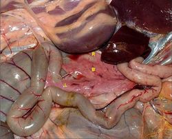
El páncreas está localizado en la parte craneodorsal del abdomen, en estrecha colaboración con el duodeno. Se puede dividir en tres partes: un cuerpo y lóbulos derecho y izquierdo. Los lóbulos son débilmente unidos por tejido conectivo interlobular. El tejido conectivo contiene vasos sanguíneos, nervios y vasos linfáticos. En general, la vena porta se extiende entre los lóbulos derecho y izquierdo (ver las species differences). El páncreas tiene más o menos una forma "V" en todas las especies. Como se mencionó en la sección del desarollo de, hay dos conductos presentes en el páncreas. Su presencia refleja el patrón de desarrollo convergente del páncreas, sin embargo, en algunas especies de uno u otro de los conductos puede atrofiar. El conducto pancreático es el más grande de los dos y se abre en el duodeno con el conducto biliar en la papila duodenal mayor. El conducto accesorio se abre en la cara opuesta del duodeno en la papila duodenal menor.
Función Exocrino
La Secreción Alcalina
El jugo pancreático vierte en el duodeno a través de conductos. Es alcalina, ya que contiene bicarbonato y los iones de cloruro. Los iones de bicarbonato son transportados activamente al lumen del conducto. El agua sigue pasivamente por ósmosis. La osmolaridad del jugo pancreático es equivalente a la osmolaridad de la sangre. El jugo pancreático es alcalino para neutralizar el jugo gástrico ácido. Esto es ventajoso porque: proporciona un pH óptimo de las enzimas del páncreas y evita daños a la mucosa delgada de absorción del duodeno. Su alcalinidad también ayuda a amortiguar el intestino grueso que es importante en fermentadores del intestino grueso.
La Digestión Enzimática
La secreción alcalina contiene enzimas digestivas que pueden digerir las proteínas, carbohidratos y lípidos. La digestión se discute en el resumen del intestino delgado.
La tabla siguiente se muestra las enzimas segregadas por el páncreas y sus efectos:
| Enzima | Sustrato | Acción |
|---|---|---|
| Tripsina, quimotripsina, elastasa | Péptidos | Endopeptidasas; corte los bonos unirá entre los aminoácidos |
| Carboxipeptidasa y aminopeptidasa | Péptidos | Exopeptidasas; corte los bonos en el término de un péptido |
| α - amilasa | Polisacáridos: almidón y glucógeno | Endoglycosidase; corte los bonos entre monómeros de los hidratos de carbono para producir la maltosa y las cadenas cortas de hidratos de carbono. |
| Lipasa pancreática | Triglicéridos y de 1,2 - diacilgliceroles | Ácidos grasos, glicerina y 2 - monoacilglicerol |
Función Endocrino
Las unidades funcionales son los islotes de Langerhans, que están incorporadas en todo el tejido exocrino. Las células de los islotes producen hormonas que mantienen la normoglucemia. El glucagón eleva el nivel de glucosa en la sangre y la insulina decrece el nivel de glucosa en la sangre. La somatostatina actúa de manera paracrina para inhibir la secreción de insulina y glucagón. Se piensa que polipéptido pancreático es un antagonista de la colecistoquinina, y por lo tanto inhibe la secreción del jugo pancreático.
Ver aquí para más información sobre el Glucagón y la Insulina.
Diabetes Mellitus
Una condición que se caracteriza por la incapacidad de mantener la normoglucemia, con hiperglucemia persistente observada. Los signos clínicos incluyen: glucosuria, poliuria y polidipsia - la concentración de glucosa en la sangre excede el umbral renal (~10 mmol/l) y se excreta en la orina. El mayor potencial osmótico del filtrado se extrae a agua al filtrado que se pierde en la orina. El animal bebe más para compensar la pérdida de agua. Polifagia y pérdida de peso también se observan cuando el animal compensa la pérdida persistente de la glucosa. Cetosis y cetonuria también se ven, donde cetonas son liberados para la energía.
Tipos de Diabetes en los Perros
Deficiencia de Células β - La mayoría de los casos de diabetes mellitus en perros son de este tipo. Produce una incapacidad para producir insulina. Puede ser causada por defectos congénitos, la pancreatitis y la autoinmunidad.
El Antagonisma de la Insulina - Visto en hembras en diestro, o en animales con Cushing's (hiperadrenocorticismo). Progesterona, hormona del crecimiento y el cortisol son los antagonistas de la insulina.
Tipos de Diabetes en los Gatos
Insulin Dependant Diabetes Mellitus (IDDM); insulin deficiency - similar to human diabetes type 1. There is a failure to produce insulin. This can be caused by islet-specific amyloidosis or chronic pancreatitis leading to β cell destruction.
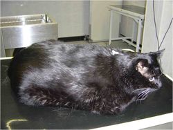
Non Insulin Dependant Diabetes Mellitus (NIDDM); insulin antagonism - similar to human diabetes type 2. Caused by obesity which leads to carbohydrate intolerance.
Vasculatura
The pancreas receives its blood supply from the coeliac and cranial mesenteric as branches from the splenic, hepatic and superior mesenteric arteries. The right lobe receives blood from the cranial pancreatoduodenal artery; a branch of the hepatic artery. The left lobe receives blood from the splenic artery and caudal pancreatoduodenal artery; a branch of the cranial mesenteric artery. Vessels aborize within the connective tissue septa and give off rich capillary networks that surround each acinus and invade each islet. The endothelium in the exocrine pancreas is continuous, whereas the endothelium of capillaries surrounding the islets in the endocrine pancreas are fenestrated. The pancreas is drained by veins that open into the portal vein. The islets receive ample blood supply to enable an appropriate response to blood glucose level.
Innervacion
The pancreas receives sympathetic and parasympathetic supply. The parasympathetic is supplied by the dorsal vagal trunk and the sympathetic is supplied by the solar plexus (splanchnic nerves). Parasympathetic and sympathetic stimulation results in exocytosis and accumulation of secretory vesicles in the lumen and ducts of the acini. Nervous regulation is thought to be of lesser importance than hormonal control. Hormones that increase pancreatic secretion include: cholecystokinin (CCK), secretin and gastrin. There is some negative feedback from somatostatin and enkephalins.
Linfáticos
Blind ending lymphatic vessels drain into larger lymphatic vessels that follow the course of the blood vessels in the connective tissue. Lymph drains into the pancreaticoduodenal lymph nodes. It then drains into the coeliac centre, which surrounds the coeliac artery.
Histología
Exocrino
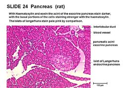
The exocrine pancreas consists of acini, which resemble bunches of grapes. Each acinus consists of a single layer of 40 - 50 pyramidal epithelial cells surrounding a lumen. The epithelial cells produce the secretion (pancreatic juice) containing enzymes, ions and water. The cells become wider during active secretion. The base of the acinar cells are strongly basophilic owing to the presence of endoplasmic reticulum, where there is a high concentration of RNA. This part of the cell therefore stains darker with haematoxylin and eosin. The apex of the cells is abundant with secretory granules containing the zymogen precursors of the pancreatic enzymes. The number of secretory granules increases after fasting, and decreases after a meal.
The lumen of the acini drain into the intercalated duct. Intercalated ducts converge to make larger interlobular ducts, which in turn converge to make interlobar ducts. Interlobar ducts are found in the connective tissue septa between lobules. Interlobar ducts join to form either the pancreatic or the accessory duct, these ducts drain into the duodenum. In some cases, the pancreatic duct unites with the bile duct, and bile and pancreatic juice enter the duodenum together.
Endocrino
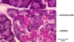
In the endocrine pancreas, the islets of Langerhans are embedded in the exocrine tissue. Each islet is composed of 2 - 3 thousand epithelial cells. The epithelial cells are arranged in a compact structure that is pervaded by a capillary network. A thin layer of reticular fibres separates the islets from the surrounding exocrine tissue. There are four different cell types within the islets of Langerhans that each produce different hormones, they include:
α cells- Produce glucagon, typically located at the periphery of the islet. They are not present in all islets.
β cells- Produce insulin. The predominant cell type, located in the centre of the islet and contributing to 70% of all cells.
δ cells- Produce somatostatin. There are low numbers in all islets.
F cells- Produce pancreatic polypeptide and are few in number, they may be present in the exocrine tissue also.
Diferencias Entre las Especies
Carnivore
Carnivores have a pancreas that is clearly distinguishable as a body and left and right lobes. The portal vein runs dorsally between the left and right lobes. The left lobe is smaller than the right. The tip of the left lobe contacts the left kidney and lies in the greater omentum. The right lobe follows the descending duodenum and lies in the mesoduodenum. Dorsally, it is related to the visceral surface of the hígado and the ventral surface of the right kidney. Ventrally, it is related to the descending duodeno. Laterally it is related to the ascending colon. In dogs, both pancreatic and accessory ducts persist throughout development. However, the pancreatic duct is smaller. It joins the bile duct just before opening into the major duodenal papilla which lies 3-6cm distal to the pylorus of the estómago. The accessory duct is the bigger duct and opens 3-5cm further distally to the pancreatic duct. The two ducts communicate inside the pancreas.
In cats, the distal part of the accessory duct atrophies during development, so only the pancreatic duct persists. Cats normally have pacinian corpuscles in the interlobular tissue that are visible grossly being 1-3cm in diameter. Dogs and cats produce little pancreatic juice between meals, but lots during a meal.
Rumiantes
The pancreas of a ruminant consists of a distinguishable short body and left and right lobes. The left lobe lies in the retroperitoneal space and is in contact with the liver, diaphragm and major vessels dorsally. Ventrally, it is in contact with intestines and dorsal sac of the rumen. The right lobe is larger and lies in the mesoduodenum against the flank of the animal and runs part of the length of the descending duodenum. The portal vein passes dorsally at the pancreatic notch between the left and right lobes. In the ox, the distal part of the pancreatic duct atrophies during development, so only the accessory duct persists. The accessory duct enters the duodenum 20 to 25cm distal to the entry of the bile duct. In sheep and goats the distal part of the accessory duct atrophies during development, so only the pancreatic duct persists. It unifies with the bile duct so that both enter via a common duct. There is a constant secretion of pancreatic juice.
Equina
The pancreas lies mainly on the right, in the very dorsal part of the abdomen. It is triangular in shape and lies within the sigmoid flexure of the duodeno. The lobes are less distinguishable compared to the dog. The ventral surface is directly attached to the right dorsal colon and base of the ciego. The dorsal surface is directly attached to the right kidney and hígado. The portal vein perforates the pancreas at the pancreatic ring. Both the pancreatic and accessory ducts persist throughout development. There is a constant secretion of pancreatic juice, which increases after feeding. This provides the caecum and colon with a constant supply of buffered solution, which maintains a stable environment important for microbe survival.
Porcino
The pancreas consists of a large body and left lobe, with a much smaller right lobe. The portal vein perforates the pancreas. The distal part of the pancreatic duct atrophies during development, so only the accessory duct persists. Two thirds lie to the left of the midline. The right portion lies adjacent to the descending duodenum and its cranial border contacts the liver. The left portion is related to the bazo, cranial pole of the left kidney and the fundus of the estómago.
Enlaces
Comprobar tus conocimientos con los Flashcards del Páncreas
Click here for information on patología del páncreas
Enlaces de Video:


